The skull is comprised of the 14 bones of the cranium, the mandible, and the hyoid.
This page represents complete specimens of skull bones. For more examples of cranial bones, visit the Digital Teaching Collection.
The bones of the cranium articulate with each other by way of sutures, which are fibrous joints between two interlocking denticulate edges which allow very little movement. This type of joint is very different from the more familiar synovial joints, highly mobile joints encased by a sac and lubricated with fluids. Most familiar joints such as the knee, elbow, and hip are synovial joints. The only synovial joints in the skull are the jaw joint and the articulations of the tiny auditory ossicles. The sutures between bones of the cranium are most evident in young individuals. Over time the bones fuse together such that some sutures in extremely old individuals are virtually invisible.
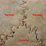
Lambdoidal suture detail
Most of the sutures are named based on the bones involved (e.g. the frontonasal sutures). However, several of the sutures have special names. The coronal suture is between the frontals and parietals, the sagittal suture is where the two parietals meet, the squamosal suture is at the junction between the temporal and the parietals, and the lambdoidal suture is at the union of the parietals and occipital.
For other on the 14 bones that make up the cranium, visit eSkeletons.
Certain aspects of the cranium can be used to estimate ancestry.
The mandible consists of a horizontal portion, the corpus, and a vertical portion, the ascending ramus. The corpus bears alveoli which anchor the teeth in place. At midline where the two hemimandibles are joined the mental protuberance, or the chin, projects anteriorly from the inferior margin of the external surface of the corpus. Just superior and posterior to the chin lies the mental foramen. The interior surface of the corpus bears several mental spines near the midline. Just posterior to the spines, the mylohyoid line courses obliquely from inferior to superior. This line provides the attachment for the mylohyoid muscle.
The angle where the ramus meets the corpus is termed the gonial angle. The edge of this angle bears the roughened masseteric tuberosity, the insertion site of the masseter muscle. The ramus rises superiorly and ends in a condyle situated posterior to the coronoid process. The process is separated from the condyle by the mandibular notch. The condyle forms the lower half of the joint whereby the mandible articulates with the glenoid fossa of the temporal bone. The variably shaped coranoid process provides the insertion point for the temporalis muscle. The interior surface of the ramus is decorated by the mandibular foramen, a large hole which pierces the ramus obliquely. This foramen transmits the inferior alveolar nerve and vessels.