FORENSICS

  • Learning Modules
    • Age Estimation
    • Ancestry
      • Ancestry: Cranium
      • Ancestry: Dentition
    • Sex Determination
    • Pathology
    • Stature Estimation
    • Taphonomy
      • Animal Activity
      • Burial Damage
      • Fire Damage
      • Mineral Absorption
      • Weathering
  • Case Files
  • Glossary
  • News
  • FAQ

Osteology: Carpals

July 7, 2017 By

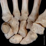
The eight carpal bones are small, compact, irregularly shaped bones arranged in two rows.  From lateral to medial, the proximal row consists of the scaphoid, lunate, triquetral, and pisiform.  The distal row, from lateral to medial, includes the trapezium, trapezoid, capitate, and hamate.  The trapezium, scaphoid, hamate, and pisiform each serve to anchor a fibrous band of tissue known as the flexor retinaculum.  The retinaculum forms the carpal tunnel, through which the flexor muscles pass from the lower arm to act on the hand.

Because each carpal bone has such a distinct shape, they are not easily confused with one another.  However, as the shapes are extremely irregular, they can be difficult to master.  Each carpal (except the pisiform in some cases) can be sided unambiguously by identifying the facets for other carpals and applying your knowledge of the anatomical relationships between the carpal bones.  Even so, because of the complex articulations of these bones positional siding “tricks” can be especially useful for carpals.

Return to the metacarpals or manual phalanges.


Scaphoid


Lunate


Triquetral


Pisiform


Trapezium


Trapezoid


Capitate


Hamate


The scaphoid is boat shaped, with a large concave facet for the capitate on its distal-medial margin and a large convex facet for the radius on its proximal margin.  The distal-lateral end of the scaphoid bears the scaphoid tubercule which projects in the palmar direction and anchors the flexor retinaculum.  The dorsal side of the tubercule bears another convex facet for the trapezoid and the trapezium.

Siding:  Position the bone in proximal view, with the convex facet for the radius facing you, with the tubercule pointing up.  The tubercule is on the side from which the bone comes.


As suggested by its name, the lunate is shaped like a crescent moon.  The concave facet on the distal end is for the capitate.  The capitate facet is continuous with the smaller, nearly flat facet for the triquetral on the medial side of the bone.  Just lateral to the the capitate facet is a small crescent-shaped facet for the scaphoid.

Siding:  Position the bone in a distal view, with the large crescent-shaped capitate facet facing you, with the triquetral facet pointing upwards.  The triquetral facet is on the side from which the bone comes.


The triquetral has three articular surfaces.  The isolated articular surface for the pisiform on the palmar surface of the bone is the smallest of the three articular surfaces.  The articular surfaces for the lunate (on the distal-lateral aspect) and the hamate (on the proximal-lateral aspect) form a 90° angle with each other.

Siding:  Position the bone such that the line where the lunate and hamate facets meet is vertical, and the pisiform facet is up.  The pisiform facet will be on the side from which the bone comes.

The smallest of the carpal bones (actually a sesamoid bone), the pisiform is a pea-shaped bone bearing only a single articular facet for the triquetral.  The pisiform serves as an attachment site for the flexor retinaculum.

Siding:  The pisiform cannot always be accurately assigned to side, although the lateral surface usually bears a small grove for the ulnar artery.


The trapezium is distinctive due to its large, saddle-shaped, distally oriented articular surface for the first metacarpal and its elongate palmar ridge for the attachment of the flexor retinaculum.  The small proximal facet for the scaphoid meets the larger medial facet for the trapezoid at an obtuse angle.

Siding:  Orient the bone such that the saddle-shaped articular surface for the first metacarpal is away from you and the laterally-positioned palmar ridge is up.  This ridge will be on the side from which the bone comes.


The trapezoid is a small bone which is often described as boot-shaped.  The broad “sole” of the boot is the dorsal surface.  The bone tapers to a point on its palmar surface.  The most pointed corner of the  dorsal surface (formed by the facets for the trapezium and scaphoid) is proximal and lateral. The distal aspect of the bone bears a wedge-shaped facet for receiving the saddle-shaped base of the second metacarpal.  A small facet for the capitate is visible on the medial side of the bone.

Siding:  Orient the “boot” with the “sole” down with the V-shaped junction of the second metacarpal and trapezium facets facing you.  The boot will point to the side from which the bone comes.

The capitate is the largest of the carpals.  It has a rounded head on its proximal end which articulates with the concave portions of the scaphoid and lunate.  Distally the capitate has a broad base which articulates primarily with the third metacarpal, though there are smaller articulations with the second and sometimes fourth metacarpals.  The medial side bears an articular surface for the hamate which runs the full proximal-distal length of the the bone.  The lateral side bears a much smaller facet for the trapezoid.  The dorsal and palmar surfaces of the bone are non-articular.

Siding:  Consider that the head of the capitate is proximal, and the dorsal surface is much broader and flatter than the ventral surface.  The dorsal surface bears a large pointed process which points medially.  If you position the bone so that the broad dorsal surface is down, and the base for the metacarpal articulations is facing you, the pointed process will be on the side from which the bone comes.

The hamate is distinctive due to its hook-shaped hamulus, which projects from the proximal-medial corner of the palmar surface of the bone.  The hamulus is an attachment site for the flexor retinaculum. The proximal end of the hamate bears a slightly convex, broad articular surface for the fourth and fifth metacarpals.    The lateral surface bears a large articular surface for the capitate, while the medial surface has an irregularly shaped facet for the triquetral.

Siding:  Position the bone such that the hamate is up, and the metacarpal articular surfaces are away from you.  The hook of the hamate leans towards the side from which the bone is from.


0.00 cm

0%

©eForensics 2025

Acknowledgements

Department of anthropology

University of Texas at Austin

Liberal Arts Instructional Technology Services

Production Credits

About

eForensics provides interactive environment using real cold cases as examples for forensic anthropology techniques. If you have problems using this site, or have other questions, please feel free to contact us.

Images and content are created by faculty, staff, and students at the University of Texas. All photographs, images, and text are copyrighted by eForensics, John Kappelman, or the University of Texas at Austin, and licensed by Creative Commons. Reproduction or recreation is permitted under not for profit circumstances and enterprises with proper attribution to eSkeletons.org.

eAnthro Digital Laboratories

3D Printing

eANTHRO LABS

eFOSSILS

eSKELETONS

eLUCY